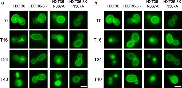
Fig. 3.

Membrane localization of Hxt36 and Hxt36 N367A fused to GFP with and without the 3K (K12,25,56R) mutations when grown on minimal medium with 2 % d-glucose (a) and 2 % d-xylose (b) in a 0–40 h time range. The scale bar corresponds to 5 μm

Membrane localization of Hxt36 and Hxt36 N367A fused to GFP with and without the 3K (K12,25,56R) mutations when grown on minimal medium with 2 % d-glucose (a) and 2 % d-xylose (b) in a 0–40 h time range. The scale bar corresponds to 5 μm