
Fig. 1.
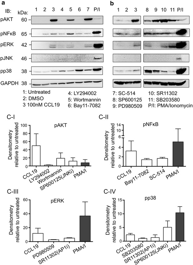
Inhibition of the CCL19-mediated signalling by pharmacological inhibitors. Resting CD4+ T cells were treated for 1 h with a predetermined (see “Methods”) concentration of inhibitor to PI3K (LY294002 and Wortmannin), NF-κB (Bay11-7082 and SC-514), JNK (SP600125), ERK (PD980509), AP-1 (SR11302) and p38 (SB203580) prior to the addition of CCL19 (100 nM) for 15 min. Cells were lysed and the level of specific phosphorylated proteins was measured using immunoblotting. a, b Representative immunoblots from two different donors treated with various inhibitors. GAPDH immunoblot was used as control for equal protein loading. c Densitometry of various phosphorylated proteins in the presence or absence of inhibitors in CCL19-treated resting CD4+ T cells. Values were normalised to both GAPDH and unactivated control. Data represent mean ± SD from 2 to 3 experiments