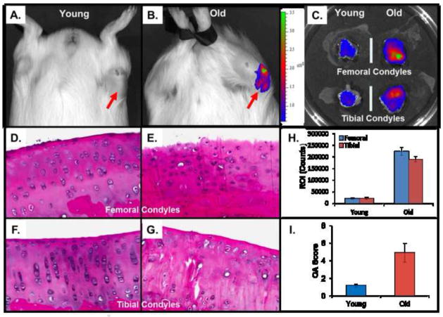
Figure 4. IVIS imaging of DH guinea pigs intravenously injected with MAbCII liposome.
Animals were IVIS imaged at 24 hours after injection. (A and B) IVIS scan images of young and older guinea pig (n=12 for each age group). (C) IVIS imaging of cartilages from dissected knee joints; (H) total integrated intensity of fluorescence (ROI) in femoral and tibial condyles was quantitated and normalized using imaging software. (D, E, F and G) Histopathology of Hematoxylin and Eosin (H&E) stained sections of articular knee cartilage from young & old GP showing regional variation in morphology. Tibial condyles were decalcified, embedded and 60 H&E-stained sections from each specimen were prepared with a constant interval of 100 μm to survey the articular cartilage. (I) These sections were then graded by an observer using a modified Mankin scale (normal; grade 0–2, slight; grade 3–5, moderate; grade 6–8 and severe; grade 9–11).

