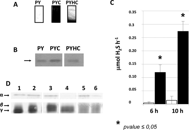
FIG 4.
Hydrogen sulfide production in strain 630Δerm. (A) Detection of H2S production using lead-acetate paper. H2S production was evaluated in the media PY, PY plus cysteine (PYC), and PY plus homocysteine (PYHC). The production of H2S yielded a black color due to the formation of PbS. (B) Detection of the homocysteine γ-lyase activity on a zymogram. Crude extracts of strain 630Δerm grown in PY, PYC, or PYHC were loaded on a native polyacrylamide gel (12%) and incubated with 10 mM homocysteine. Homocysteine γ-lyase was detected by the formation of insoluble PbS via the release of H2S. Lanes of the zymogram have been reorganized from the same image to present data chronologically. (C) Quantitative detection of H2S after 6 or 10 h of growth of strain 630Δerm in PY (white boxes) or PYC (black boxes). H2S production was measured using the quantitative methylene blue method, as described in Materials and Methods. The statistical analysis was performed by using the Mann-Whitney test for all genes. (D) Detection of cysteine desulfhydrase activities on a zymogram. Crude extracts of strain 630Δerm (lanes 1 and 2), 630Δerm::cysK (lane 3), 630Δerm::sigL (lane 4), 630Δerm(pRPF185) (lane 5), and 630Δerm(pDIA6456-ASmalY) (lane 6). The strains were grown in PY (lane 1) or PYC (lanes 2 to 6). Samples were charged on a native polyacrylamide gel (12%) and incubated with 10 mM cysteine. The cysteine desulfhydrases were detected by the formation of insoluble PbS formed by the release of H2S. The results presented are representative of at least three independent experiments. Lanes of the zymogram have been reorganized from the same image to present data chronologically.

