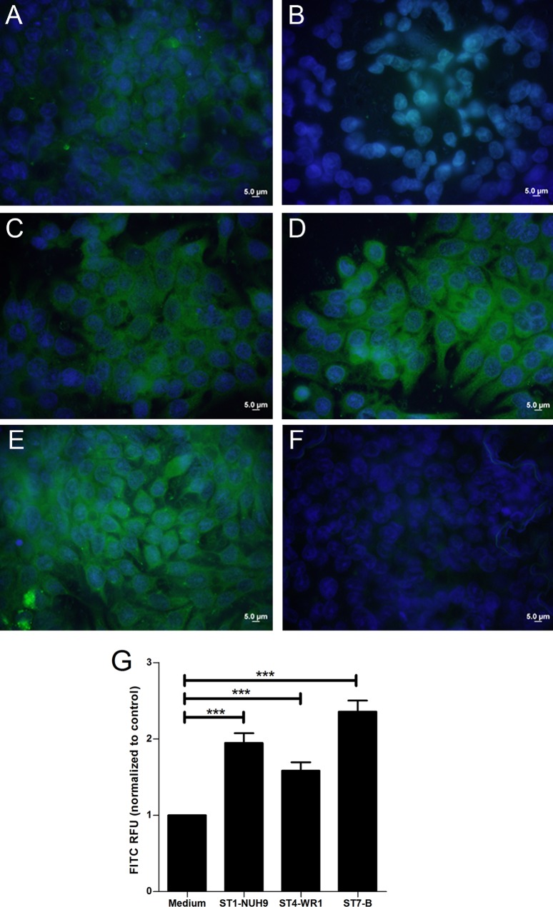
FIG 2.
Confluent HT-29 cells in monolayers increase the production of cathelicidin after incubation with Blastocystis. (A and B) Immunofluorescence assay images show a basal level of cathelicidin production in differentiated HT-29 cells (A), which was absent in undifferentiated cells (B). (B to E) Differentiated HT-29 cells incubated with Blastocystis ST1-NUH9 (C), ST4-WR1 (D), and ST7-B (E) show brighter fluorescence after 1 h of coincubation at an MOI of 10. (F) Negative control. (G) Total fluorescence units were quantified by using ImageJ software. RFU, relative fluorescence units. ***, P < 0.0001.

