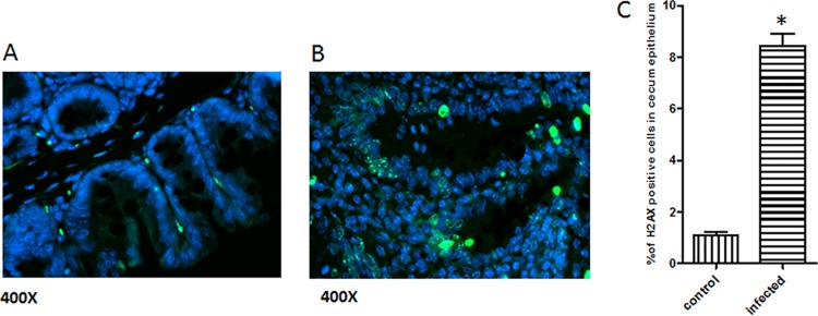
FIG 7.
Immunofluorescence staining of gnotobiotic mouse cecum for γ-H2AX (in green). Five mice in each group were examined, with the following results: the uninfected tissue had very few positively stained cells in the cecum (A) and more γ-H2AX-positive cells were observed in the epithelium, particularly within areas of intestinal hyperplasia and dysplasia in the infected cecum (B). (C) The number of γ-H2AX-positive cells in the cecal epithelium of H. saguini-infected mice was statistically higher than that in the control. (*, P < 0.05).

