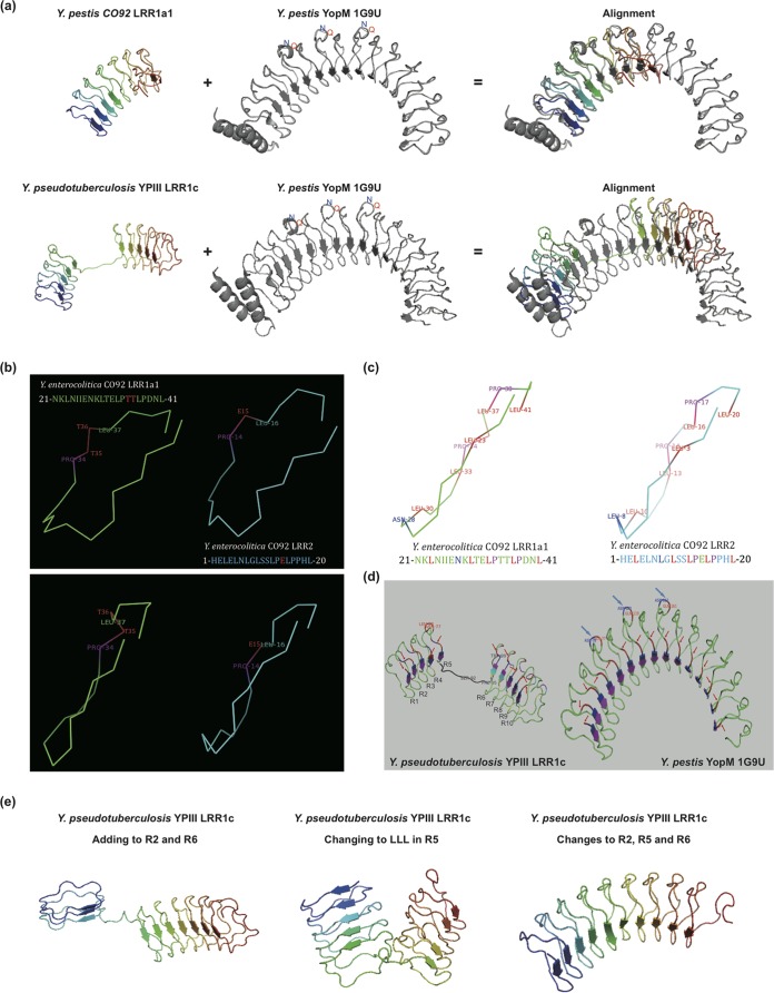
FIG 6.
LRR protein structure comparison. (a) Structural comparison of Y. pestis CO92 LRR1a1 protein or Y. pseudotuberculosis YPIII LRR1c protein and the reference structure 1G9U for Y. pestis YopM. (b) Structural comparison of the repeat units of LRR1 and LRR2 proteins. The top and side faces of the units are shown in the upper and lower panels, respectively. The key position difference is shown in red. The Y. enterocolitica CO92 LRR1a1 and LRR2 repeat units are used as representatives. (c) Shape-maintaining residues of repeat units (indicated in red, purple, and blue). (d) Repeat sequence starting difference in Y. pseudotuberculosis YPIII LLR1c protein. Each repeat sequence start is indicated with a red arrow; QN loops are indicated with blue arrows. Repeat sequence starts of the reference YopM structure (1G9U) are also shown. (e) Structure of Y. pseudotuberculosis YPIII LRR1c with the gaps in repeats R2 and R6 filled with D residues (left), with the three residues (S, F, and W) in R5 consensus positions replaced by L residues (middle), and with both the gaps in repeats R2 and R6 filled with D residues and the three residues (S, F, and W) in R5 consensus positions replaced by L residues (right).

