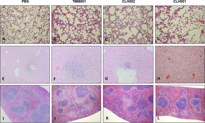
FIG 2.
Representative images of organ pathology in challenged mice. Hematoxylin-and-eosin-stained tissues displayed the types of pathology seen in lungs (A to D), livers (E to H), and spleens (I to L) of mice challenged with PBS or 1.5 × 10⁴ CFU of TMM001, CLH002, or CLH001 at 21 days postchallenge.

