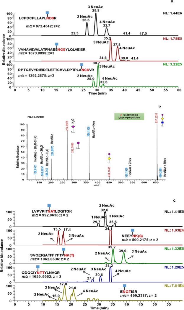
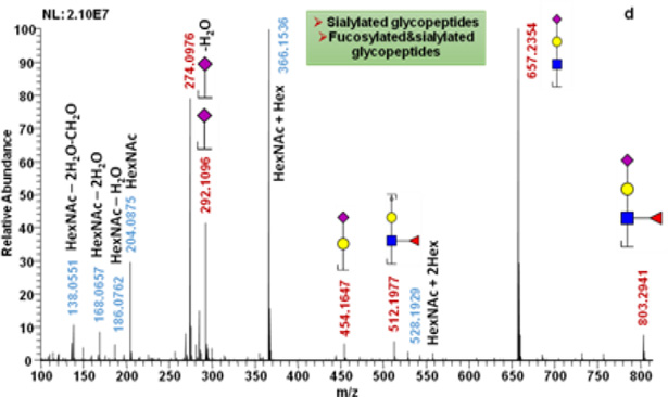
Figure 2.
Extracted ion chromatograms of Y1 ions of fetuin (a) and AGP (c) glycopeptides. Mass Spectra of the low m/z regions of the SID full MS scan of fetuin (b) and AGP (d) samples. 2, 3 and 4 NeuAC labeling is used to indicate that the detected Y1 originated from glycopeptides with glycan structures containing 2, 3 and 4 sialic acid moieties, respectively.


