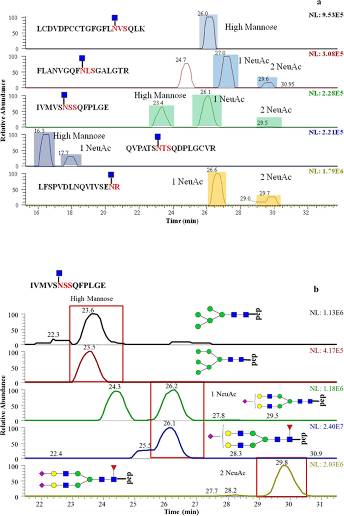
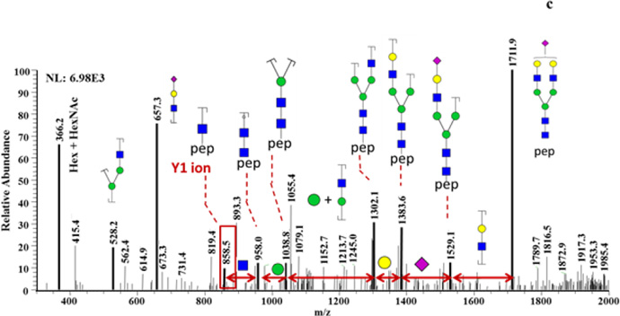
Figure 4.
PTG glycosylation sites and glycan compositions related to such sites. (a) EIC of detected Y1 ions. (b) EIC of glycopeptides associated with IVMSNSSQFPLGE peptide backbone of. (c) Tandem mass spectra of a glycopeptide that consists of peptide backbone IVMSNSSQFPLGE and HexNAc4Hex5 glycan composition. 2 and 3 NeuAc labeling is used to indicate that the detected Y1 originated from glycopeptides with glycan structures containing 2 and 3 sialic acid moieties, respectively.


