Figure 4.
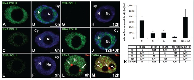
Localization of RNA polymerase II with a phosphorylated serine 2 in the CTD domain (elongation form). The roots of Lupinus luteus were imaged before (A, B) and after 6 h (C, D), 9 h (E, F), and 12 h (G, H) submerged and after 3 h reoxygenation (I, J). Decreased signal was observed in successive hours of hypoxia (C–H). After reoxygenation, the signal reappears in the nucleus (I, J). Quantitative analysis of RNA polymerase II elongation form in the nucleus (K) of meristematic root cells under normoxia, hypoxia and re-aeration conditions. The tables under plot present the significant differences between groups, with testing probability below the value shown in the table (P<), α = 0.05. In the column heading, the number of analyzed cells in each stage is presented in brackets. ns - nonsignificant differences between groups. Double immunolocalization in the nuclei cells before (L) and after 12 h hypoxia (M) of PANA antigen (green) and Sm proteins (red). The few large round speckles (arrowheads) not colocalized with Cajal bodies (arrows) occurred in the nucleus after 12 h hypoxia (M). Merging with DAPI staining (B, D, F, H, J). N- nucleus, Nu- nucleolus, Cy- cytoplasm.

