Fig. 1.

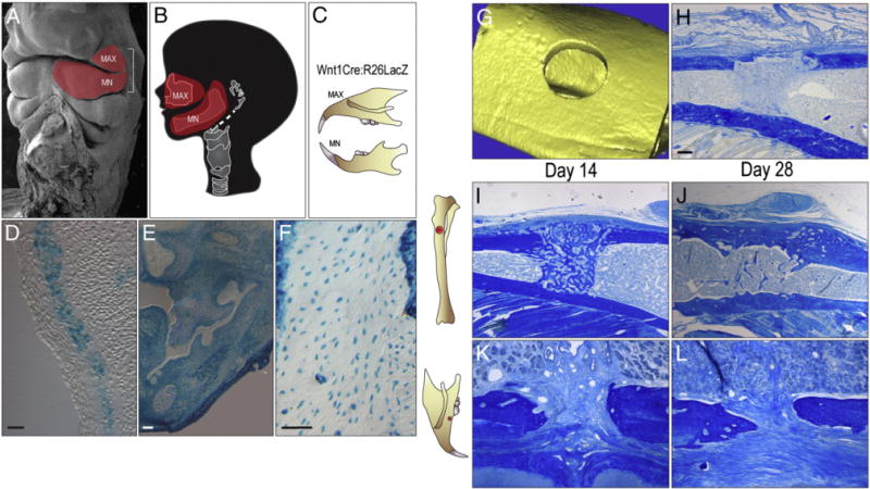
Robust bone repair and implant osseointegration in the tibia compared to the maxilla. (A) Scanning electron micrograph of embryonic head; the red region indicates cranial neural crest cells of the first branchial arch, (B) that later develop into the maxilla and the mandible. (C) The cranial neural crest cells of Wnt1Cre:R26RLacZ mice are permanently labeled. (D) Permanently labeled LacZ + cranial neural crest cells are found within the first branchial arch along with (E) many dental tissues. (F) Mature osteocytes of the maxilla are also permanently marked for their cranial neural crest origin. (G) Micro-CT image of 0.8 mm skeletal defect created in mesoderm-derived long bone, the tibia. (H) Representative tissue section through a 0.8 mm tibial defect following injury, stained with aniline blue. (I) Representative tissue section through a 0.8 mm tibial defect on post-surgery day 14, stained with aniline blue. The dark blue color denotes collagen-rich matrix in the defect site. (J) Representative section on post-surgery day 28 from the tibia implant site. (K) Representative tissue section through a 0.6 mm maxillary defect on post-surgery day 14, stained with aniline blue. The dark blue color indicates the collagen-rich matrix of the bone. (L) Aniline blue stain of the maxillary defect on post-surgery day 28. MAX, maxilla; MN, mandible. Scale bars: (D–F), 100 μm; (H–L) 100 μm.
