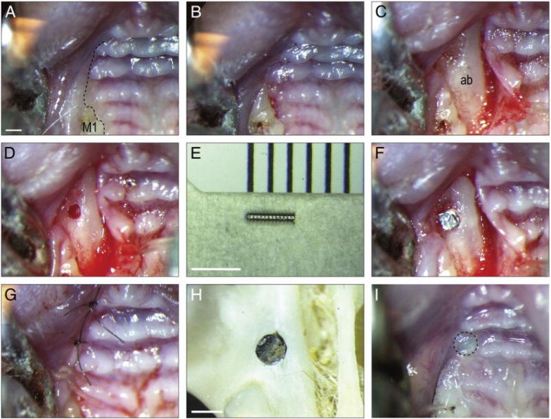
Fig. 2.

An oral implant model in mice. (A) Pre-operative photograph of the alveolar crest, anterior to the maxillary first molar; black dotted line indicates incision placement. (B) The intrasulcular incision extends from the lingual surface of the maxillary first molar anteriorly, to the crest of the edentulous space. (C) A full-thickness flap is elevated to expose the alveolar bone. (D) A 0.45 mm hole is prepared on the crest, 1.5 mm anterior of the first maxillary molar. (E) The 0.6 mm diameter titanium alloy implant. (F) The implant is placed manually, followed by careful rinsing. (G) Wounds are closed with non-absorbable single interrupted sutures. (H) Skeletal preparation showing location of the maxillary implant relative to the dentition and bones of the skull. (I) Soft tissue covered the healing implant days post-surgery. M1, maxilla first molar; ab, alveolar bone. Scale bars: (A–D, G, I) 600 μm; (E) 2500 μm; (H) 500 μm.
