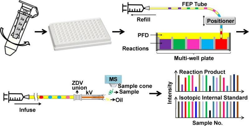
Fig. 1.

Diagram of SIRT1 assay: SIRT1 was dialyzed from Tris buffer into formate buffer using a centrifugal dialysis unit; the deacetylation reactions were conducted in formate buffer in a multi-well plate; reaction mixtures were reformatted into oil-segmented droplets in a piece of fluorinated ethylene propylene (FEP) tubing; finally, droplets were infused into an orthogonal ESI source through a modified ESI needle. The FEP tube and the needle were connected by a zero dead volume (ZDV) union. The signal intensity of the reaction product and its isotopic internal standard were monitored. Oil segment did not generate ESI signal thus showed as spacing between sample droplets
