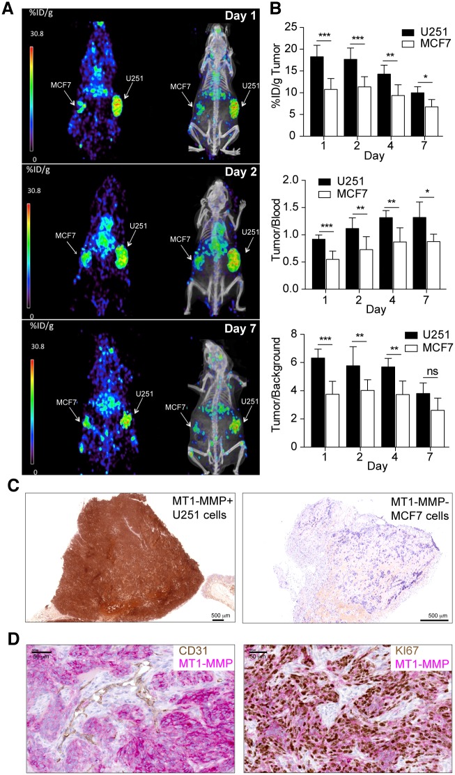
Fig 3. PET/CT imaging with radiolabeled 89Zr-DFO-LEM 2/15 in mice bearing MT1-MMP+ GBM cells (U251) and MT1-MMP- breast cancer cells (MCF-7).
A) Representative coronal whole-body PET and CT sections at 1, 2 and 7 days p.i.; B) Levels of radioactivity in tumors, tumor to blood and tumor to background ratios derived from PET imaging after 89Zr-DFO-LEM 2/15 administration to mice bearing MT1-MMP+ and MT1-MMP- tumors (mean±SD, n = 11−4/time); C) Immunohistochemistry of tumor tissue from xenografted mice used for PET imaging. MT1-MMP was detected using LEM2/15 antibody. Scale bars: 500 μm D) Representative images of double immunostaining for MT1-MMP (pink) and CD31 vascular marker (brown) (left panel) and for MT1-MMP (pink) and Ki67 proliferation marker (brown)(right panel) in U251 tumor implants from xenografted mice used for PET imaging. Scale bars: 50 μm. Significant differences: p<0.05 (*), p<0.001 (**) and p<0.0001 (***), ns, not significant.

