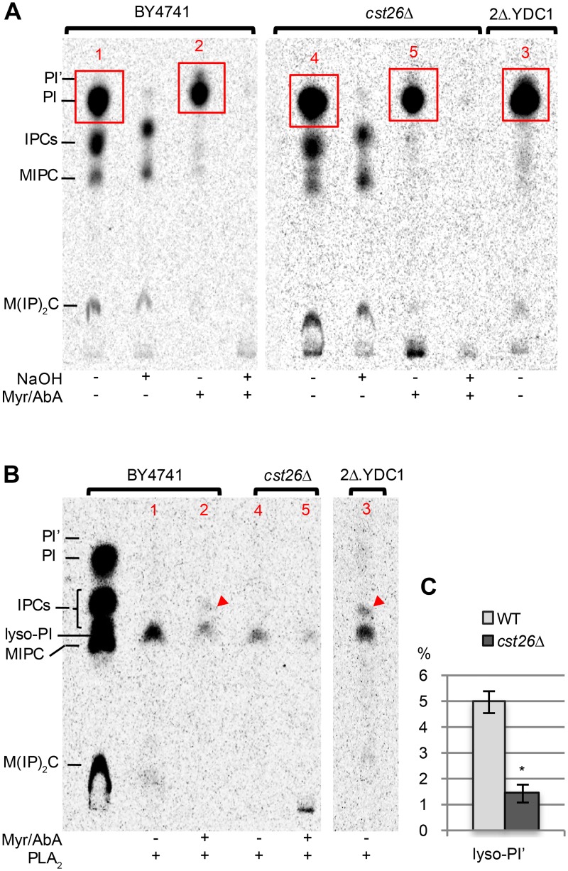
Fig 9. Cst26 introduces VLCFAs into lyso-PI.
Cells were grown to exponential phase in SC (2 mg/L of inositol). (A) 3 OD600 units of cells were washed and preincubated in inositol-free SC containing or not Myr (40 μg/ml) and AbA (1 μg/ml) for 10 min and then labeled with 10 μCi [2-3H]-myo-inositol for 4 h at 30°C. Lipids were extracted, deacylated or not with NaOH, desalted and run on TLC in solvent 1. (B) zones in red rectangles in panel A were scraped off the plate, eluted with organic solvent, dried in a rotary evaporator and treated with PLA2. Red arrowheads point lyso-PI’. (C) radioscanning was used to quantify radioactivity in lanes containing lipids from Myr/AbA treated cells. Radioactivity was measured in the zone where lyso-PI’ migrates and also in the rest of the lane (except for the origin); lyso-PI’ was then calculated as a percentage of total radioactivity in the lane. In this way, results of the experiment of panel B and a second identical experiment were averaged. * P = 0.002.

