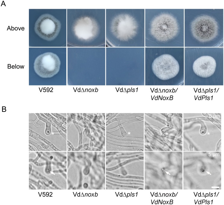
Fig 2. Penetration assay on the cellophane membrane.
A. Colonies of V592, VdΔnoxb and VdΔpls1 mutant strains, VdΔnoxb/VdNoxB and VdΔpls1/VdPls1 complemented strains grown on MM medium overlaid with a cellophane layer (above) and removal of the cellophane membrane (below). Photographs in the first row were taken at 7 dpi. The second row shows growth of a V592 colony on MM medium after penetration from the cellophane membrane; neither mutant strain breached the cellophane membrane to grow on medium. Complemented strains restore the penetration ability. B. Observation of penetration peg development on the cellophane membrane at 2 dpi. Differentiation of hyphopodia (swollen hyphae) in V592, mutant and complemented strains was indicated by asterisks in the first row. Penetration pegs (the dark pin) were only observed in V592 and complemented strains from the hyphopodium indicated by the arrow in the second row, which was focused at 5μm below the first row. Bar = 5μm.

