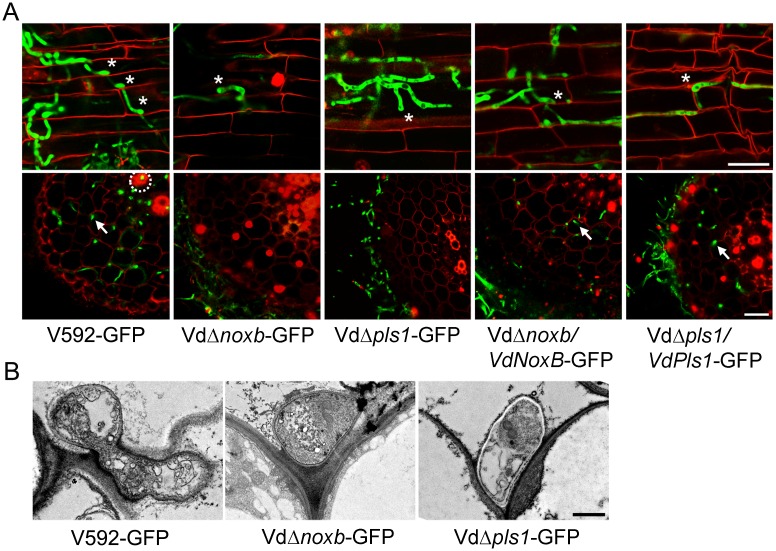
Fig 3. Observation of fungal penetration in cotton roots.
A. Confocal laser scanning microscopy (CLSM) images of hyphopodium development and/or penetration of V592-GFP, mutant strains VdΔnoxb-GFP and VdΔpls1-GFP and complemented strains VdΔnoxb/VdNoxB-GFP and VdΔpls1/VdPls-GFP. Photographs were taken on the surface (first row) or the cross section (second row) of cotton root at 3dpi and 5dpi, respectively. Asterisks indicate the hyphopodium. Root xylem structure was labeled by white dotted line, and invasive hyphae were indicated with arrows. Bar = 25 μm. B. Transmission electron microscope (TEM) images show penetration of a V592-GFP hyphopodium, but not those of the VdΔnoxb-GFP or VdΔpls1-GFP mutants, through the root cell wall. Bar = 1 μm.

