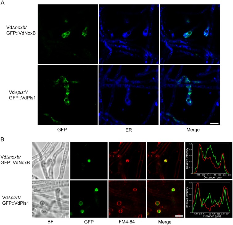
Fig 4. CLSM observation of cellular localization of VdNoxB and VdPls1.
A. Localization of GFP::VdNoxB and GFP::VdPls1 in hyphopodia at small vesicles and the ER. VdΔnoxb-GFP::VdNoxB and VdΔpls1-GFP::VdPls1 strains were inoculated on MM medium overlaid with cellophane membrane. Photographs were taken at 2 dpi. ER was stained by ER-Tracker Blue-White DPX. B. CLSM observation focused at the base of hyphopodium and corresponding linescan graphs. Co-localization of either GFP::VdNoxB- or GFP::VdPls1 with plasma membrane stained with FM4-64. Linescan graphs showing either VdNoxB or VdPls1 localizes on the plasma membrane in a transverse section of individual hyphopodium. Bar = 5 μm.

