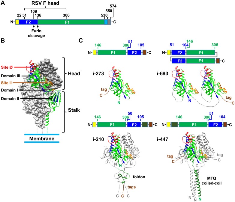
Fig 1. Design of RSV F head-only immunogens.
(A) Genetic construct of RSV F illustrating the location of the head region. The signal peptide is colored yellow, F2 is colored blue, F1 is colored green, the transmembrane region is colored cyan and the cytoplasmic domain is colored gray. (B) Pre-F form of the RSV F trimer. One protomer of RSV F is depicted as a ribbon diagram and colored as in A. The other two protomers are gray surface representations. Antigenic site Ø and site II are red and orange respectively. Domains I, II and III are labeled as defined previously [9, 35]. (C) Models of head-only immunogens i-273, i-693 (upper panels), i-210 and i-447 (lower panels). For each immunogen a cartoon of the genetic construct is depicted (top) and a ribbon diagram (bottom), color-coded as in A and B with brown coloring for purification tags. PDB entries 4JHW [9], 1RFO [36] and 1GCM [37] were used to depict RSV F, the foldon and the coiled coil respectively. RSV F residue numbering follows the numbering in PDB entry 4JHW [9].

