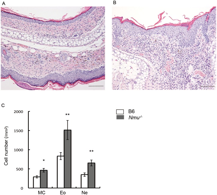
Fig 3. Histological analyses of the ears of B6 and Nmu-/- mice after repeated hapten exposure.
(A) Hematoxylin-eosin (HE) staining of B6 and (B) Nmu-/- mice on day 28 (n = 5). Original magnification, ×100. (C), Numbers of histologically identifiable dermal mast cells (MC), eosinophils (Eo), and neutrophils (Ne) in the ears of B6 and Nmu-/- mice (n = 5). *P < 0.05, **P < 0.01.

