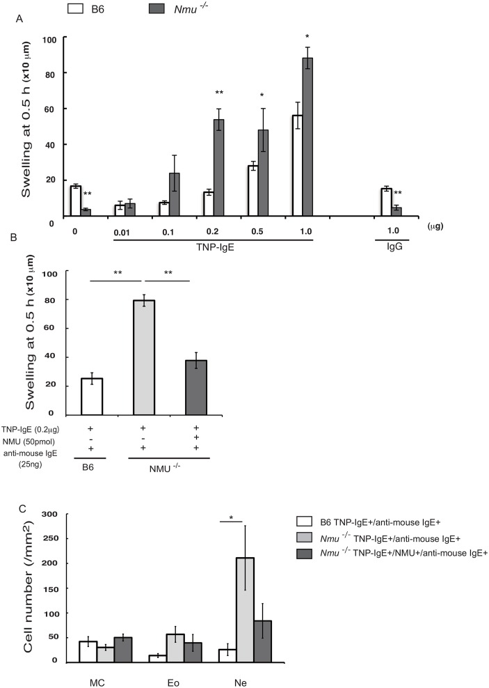
Fig 6. Effects of NMU on ITH mediated by two types of FcεRI cross-linking.
(A) ITH responses were induced by TNCB application to the footpad of B6 or Nmu-/- mice previously injected with TNP-specific IgE (S1A Fig). Different concentrations of TNP-specific IgE (0.01–1.0 μg in 20 μl of PBS per footpad) were injected 3 days before applying TNCB as follows: 0.05 μg to 0.2 μg of IgE achieved concentrations equivalent to that achieved in the serum of these mice after repeated hapten exposure on days 7–21 (2.5–10 μg/ml). (B) ITH was induced by injecting 0.2 μg of TNP-specific IgE into the footpads of B6 or Nmu-/- mice followed by the injection of 25 ng of anti-mouse IgE. NMU (50 pmol) or vehicle (PBS) was injected one day before injection of mice with an anti-mouse IgE (S1B Fig). In B6 mice, ITH was decreased by NMU released from keratinocytes upon injection compared with that of similarly treated Nmu-/- mice. (C) Numbers of histologically identifiable dermal mast cells (MC), eosinophils (Eo), and neutrophils (Ne) in the footpads of B6 and Nmu-/- mice (n = 4). Data are expressed as the mean ± SEM (n = 10), *P < 0.05, **P < 0.01.

