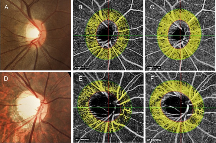
Fig 2. Example of peripapillary perfusion in the non-tessellated and tessellated eyes.
Disc photographs (A, D), the RNFL OCT angiograms (B, E) and the whole retinal OCT angiograms (C, F) in the eyes of non-tessellated group (A–C) and tessellated group (D–F). The dense microvascular network of RNFL and whole retina was lower in tessellated group than that of non-tessellated group.

