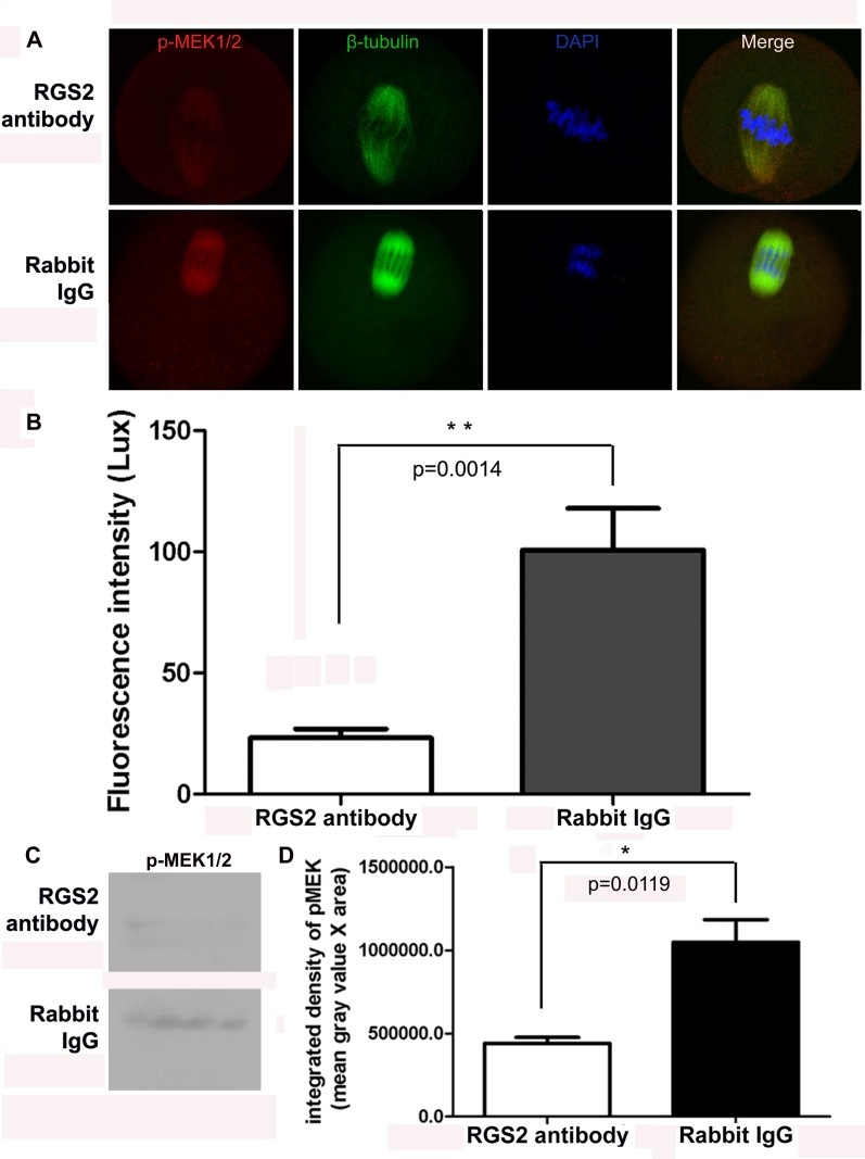
Fig 5. Antibody-mediated inhibition of the binding of RGS2 and β-tubulin reduced the expression of p-MEK1/2 in meiotic spindles during oocyte maturation.
(A) p-MEK1/2 was localized in the microtubule-organizing center of spindles in rabbit IgG-injected oocytes. However, in anti-rgs2 antibody-injected oocytes, the p-MEK1/2 signal was very week or not visible. p-MEK1/2 was labeled with Alexa Fluor 594 (red), while β-tubulin was stained with FITC (green) and nuclei were stained by DAPI (blue). (B) Fluorescence intensities of p-MEK1/2 protein in control and anti-Rgs2 antibody-injected oocytes were assessed. Images were obtained using the same laser confocal microscope using the same exposure parameters (HV 520 V; Gain X1; offset 8%). Mean fluorescence intensities were significantly different (t-test, p<0.01) between the control IgG- and anti-Rgs2 antibody-injected oocytes; 10 oocytes were measured per group. (C) Western blot analysis of p-MEK1/2 in anti-rgs2 antibody and rabbit IgG-injected oocytes. Blot is representative of 3 independent replicates; each sample includes 50 oocytes. (D) Quantitation of p-MEK1/2 blot. Data are integrated density of blot (mean gray value x blot area) ±SD from three independent experiments. Difference is considered to be significant at p<0.05.

