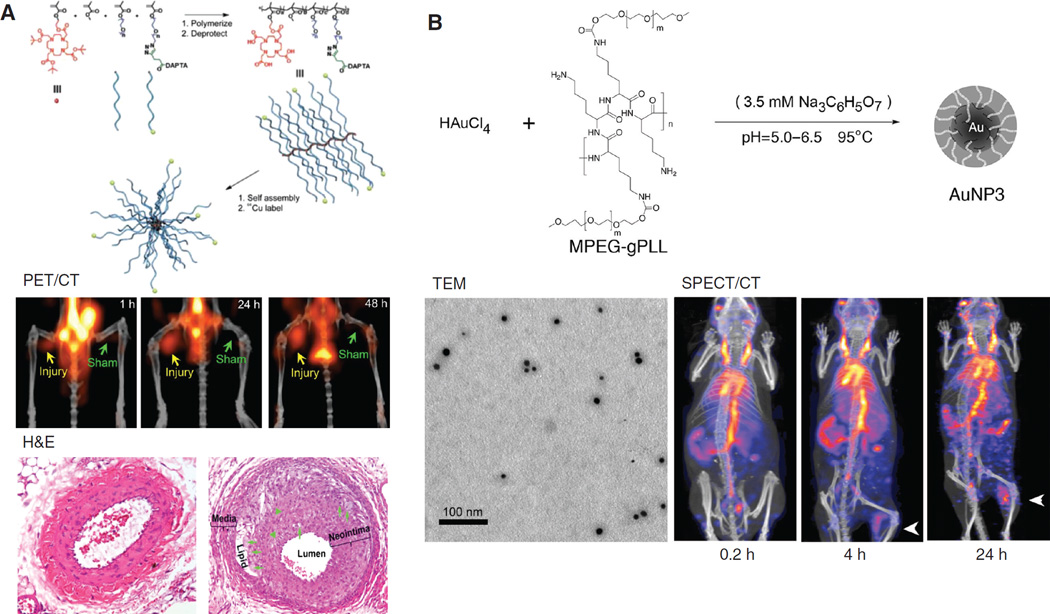
Figure 3.
Imaging of cardiovascular disease and inflammation with radio-nanomaterials. (A) Use of 64Cu-labeled, DAPTA-conjugated comb nanoparticles for imaging of wire-injury induced atherosclerosis. Significantly higher uptake of these nanoparticles was found in the injury area compared with sham-operated area. Histology examination confirmed the progressive atherosclerotic plaque in the injury group. Adapted from reference (57). (B) The structure and morphology of mPEG-gPLL grafted AuNPs and their application in detection of an experimental inflammation via SPECT/CT post 99mTc labeling. Arrows indicated the location of inflammation. Adapted from reference (58).

