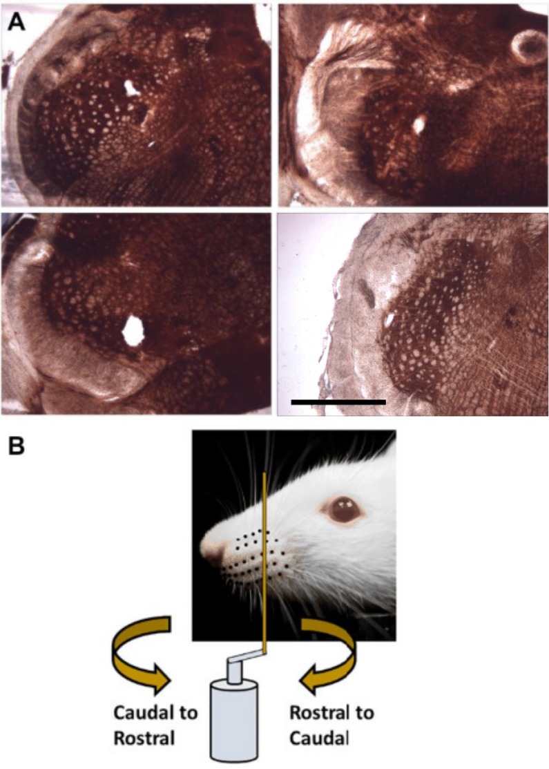
Fig 1. Recording location and whisker stimulation.
(A) Histological analysis showed lesions either in spinal trigeminal nucleus interpolaris or spinal trigeminal nucleus oralis. The scale bar represents 1 mm. (B) During stimulation a vertical metal post is mounted on a servo motor and swept through the whisker array in either Rostral-Caudal (RC; clockwise directed curved arrow) or Caudal-Rostral (CR; counter-clockwise directed curved arrow) directions and oriented perpendicular to the whisker array.

