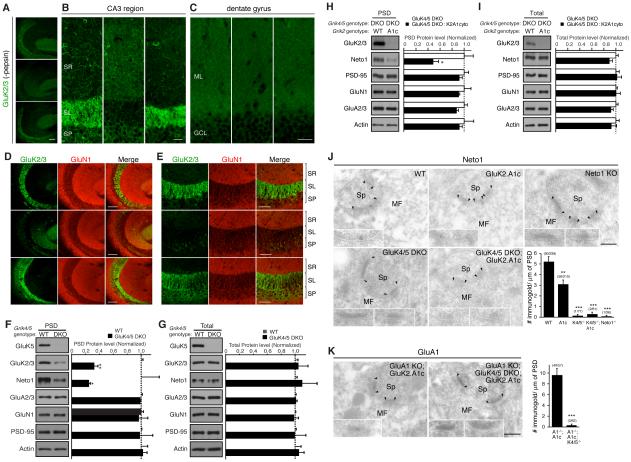
Figure 6. High-affinity GluK4/5 subunits mediate synapse specificity of KARs in the hippocampus.
The distribution of KAR components in the hippocampus was examined by immunohistochemistry and biochemical fractionation. (A and B) Immunostaining of hippocampal sections without pepsin treatment (see Experimental Procedures) revealed reduction in the GluK2/3 signal at the stratum lucidum (SL) in GluK4/5 double-knockout (DKO) mice. On the other hand, GluK2/3 signal at the stratum radiatum (SM) was elevated in GluK4/5 DKO mice. (C) GluK2/3 distribution in the dentate gyrus was unaltered (G; ML = molecular layer, GCL = granular layer). (D, E) Immunostaining of hippocampal sections after pepsin treatment. In wild-type mice (WT), a strong GluK2/3 signal was detected at the stratum lucidum, but not at the stratum radiatum or stratum pyramidale (SP). This highly compartmentalized pattern was abolished in GluK4/5 DKO mice, but was preserved in Neto1/2 DKO with a slight increase in the GluK2/3 signal at the SP. Images represent GluK2/3 localization at lower (D) and higher (E) magnifications. Scale bars: 100 μm (A, D), 50 μm (E), 25 μm (B, C). (F, G) Protein levels in the PSD fraction (F) and total (G) were measured in hippocampus (n = 5). Protein levels of KAR components (GluK2/3 and Neto1) were significantly reduced in the PSD fraction of GluK4/5 DKO, but total expression was unaltered. (H, I) Protein levels in the PSD fraction (H) and total (I) were measured in hippocampus (n = 3-4). Protein levels of KAR component (Neto1) were further reduced in the PSD fraction of GluK2.A1c KI; GluK4/5 DKO triple-mutant mice, but total expression was unaltered. (J, K) Immuno-electron microscopic images of Neto1 protein. PSDs are indicated by arrowheads. Inserts show high-magnification of labeled synapses. Scale bars = 200 nm (J) Neto1 was detected at hippocampal MF-CA3 synapses in WT mice, but not in Neto1 KO mice. On the other hand, Neto1 was reduced in GluK2.A1c KI mice. No Neto1 signal was detected in GluK4/5 DKO and GluK2.A1c KI; GluK4/5 DKO triple-mutant mice. (K) GluK2.A1cyto signal detected by anti GluA1 antibody was detected in GluK2.A1c KI; GluA1 KO double-mutant mice, but not in GluK2.A1c KI; GluK4/5 DKO, GluA1 KO quadruple-mutant mice. Numbers of immunogold-labeled synapses and total analyzed synapses are indicated in parentheses. Data are given as mean ± s.e.m. *, P < 0.05; ** P < 0.01; ***, P < 0.001 (Student’s t-test).

