
Figure 7.
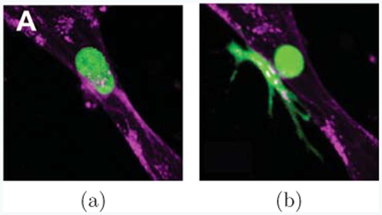
Cells can appear in vastly different shapes. Here, a cancer cell (green) extravastating from inside a vessel of endothelial cells (purple) into surrounding extracelullar matrix (black) is shown, as observed in [54] (Reproduced by permission of The Royal Society of Chemistry). In (a) the cancer cell appears nearly spherical, while it is still fully inside the vessel lumen and has not started to extravasate. When it is in the process of extravasation through the endothelium, it narrows dramatically at the endothelium, connected only through a thin neck region (b). Part of the cell remains in the lumen, but much of it has already spread outside of the lumen into the extracellular matrix.