
Figure 9.
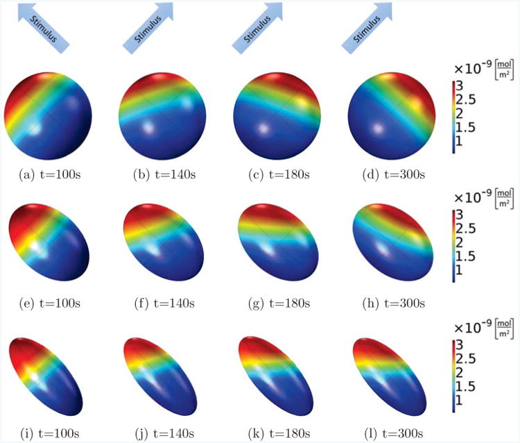
Active Rac on the membrane is shown at different times for the same cell with different shapes, where the Rac activation rate in the first 100s increases linearly along the long axis of the ellipsoid (from lower right corner to upper left corner), and from then on, it is rotated by 90 degrees and now increases linearly along a short axis of the ellipsoids (from the lower left corner to the upper right corner). In all cases, the volume of the ellipsoid cells is fixed as V = 800μm3, the main axis is 11.5μm (spherical, (a)-(d)), 15μm ((e)-(h)) and 20μm ((i)-(l)), and the other two axes are of the same length. Comparing the different shapes, we see that only the spherical cell can completely polarize into the new stimulus direction, whereas the cells with ellipsoidal shapes will form a stable pattern which points into a direction in between the original and final stimulus direction.