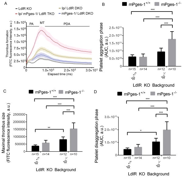
Figure 2. PGI2 restrains thrombogenesis in hyperlipidemic mice.
Thrombogenesis in cremaster arterioles after laser-induced injury in LdlR KO, Ip/ LdlR DKO, mPges-1/ LdlR DKO, and Ip/mPges-1/ LdlR TKO male mice (A). Thrombus formation was visualized in real-time with fluorescently labeled platelets as described in the Methods. Median integrated fluorescence intensity of platelets representing thrombus formation was plotted versus time after laser-induced injury of the cremaster arteriole vessel wall. Fluorescence intensity from 15–20 thrombi was averaged from each mouse. Data correspond to platelet aggregation phase (B), maximal thrombus size (C) and platelet disaggregation phase (D) were extracted from the fluorescence-time curves and averaged. Two-way ANOVA showed a significant effect of genotypes on thrombus formation in male mice aged 3–4 months (B, Ip- p< 0.0001, mPges-1- p= 0.0015, interaction- p= 0.0010; C, Ip- p< 0.0001, mPges-1- p <0.0001, interaction- p= 0.0010; D, Ip- p< 0.0001, mPges-1- p= 0.0003, interaction- p= 0.010). Holm-Sidak’s multiple comparison tests were used to test significant differences between LdlR KO and different mutants. Data are expressed as means ± SEMs. *p< 0.05, **p<0.01, ***p<0.001; n=10–15 per genotype. PA- platelet aggregation; MT- maximal thrombus; PDA- platelet disaggregation.

