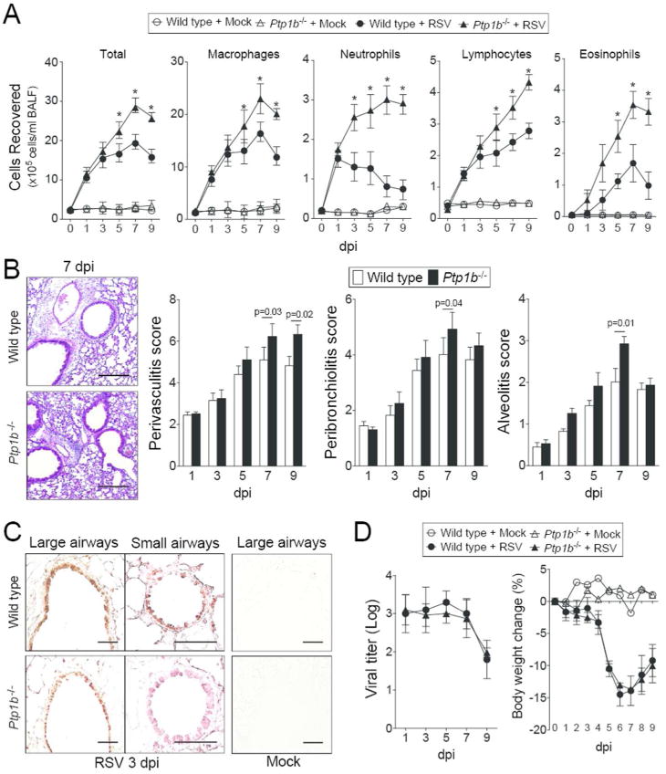
Figure 2.

Ptp1b deficient mice display enhanced lung immune cell infiltration during RSV infection. (A) Ptp1b-/- mice (triangle) and their FVB/NJ wild-type littermates (circle) were infected with 1×10⁶ pfu of RSV (closed) or mock control (open) and animals were euthanized on days 0, 1, 3, 5, 7 and 9-dpi. BALF immune cellularity was determined for total immune cell number, macrophages, neutrophils, lymphocytes and eosinophils. *Represents a p-value < 0.05 comparing Ptp1b-/- mice to wild-type mice 9 days post RSV challenge. (B) Comparative histology images of infected animals from each background 7 dpi are presented here (scale bar=50 μm). Histopathology (Peribronchiolitis, perivasculitis and alveolitis scoring) was recorded in mice for each group. Slides were randomized, read blindly and scored for each parameter. p values shown, comparing both treatments connected by a line. (C) Immunohistochemistry was performed on lung tissue from both mouse groups 3 dpi with an antibody that recognizes RSV antigen (brown). Mock treated animals demonstrate negative staining for RSV antigen. (D) RSV infectivity and animal body weight was comparable in wild type and Ptp1b-/- mice. Each graph is represented as mean ± S.E.M. where each measurement was performed on 10 animals/group, with 6 replicates/animal.
