
FIGURE 1.
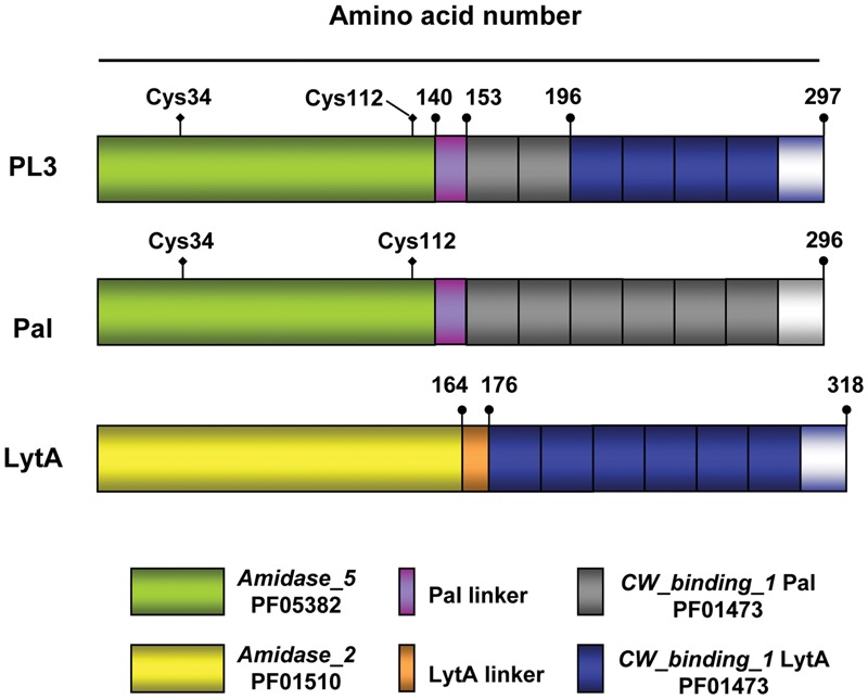
Schematic representation of the PL3 chimeric NAM-amidase and the parental Pal and LytA murein hydrolases. Domain and linker origin is depicted by colors; gray and blue rectangles indicate the choline binding repeats comprised in Pal and LytA CBDs, respectively, followed by the C-terminal tail. Numbers show the end of domains and linkers. The position of the two cysteine residues in Pal and PL3 catalytic domains is marked. Pfam entries for Amidase_2, Amidase_5, and CW_binding_1 (choline-binding repeats) families are also shown.