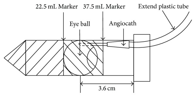
Figure 2.

Assembly of the drug distribution kit. Step 1: 22.5 mL agarose gel was poured into the 50 mL tube. Step 2: the pig eye was placed on top of the solidified agarose gel at the upright position with the angiocath connected to the extended plastic tube. Step 3: more agarose gel was poured to cover and fix the pig eye inside the 50 mL tube.
