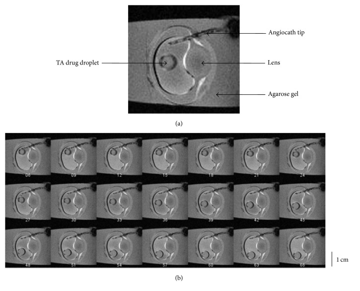
Figure 3.
Representative image series of dynamic T1-weighted MRI. (a) The angiocath tip, the drug droplet, and the pig eye lens were clearly shown in this image frame taken with MRI. (b) MR Image series showed the intraocular kinetics of a TA droplet inside the silicone oil bubble within 6 minutes after the injection. The index number under each image represents its corresponding time point after the image acquisition starts. There are in total 72 time points within each scanning session. At each time point, the T1-weighted MRI is acquired in 5 seconds.

