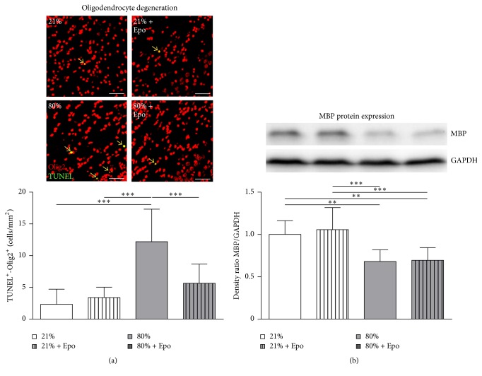
Figure 2.
Erythropoietin ameliorates oligodendrocyte degeneration but not hyperoxia-mediated hypomyelination. (a) Oligodendrocyte degeneration was determined in brain sections from P7 rats that were exposed to either normoxia (21% oxygen (21%)) or hyperoxia (24 h, 80% oxygen (80%)) at P6 and treated with normal saline or 20,000 IU/kg Epo. Oligodendrocyte degeneration was determined by immunohistochemical TUNEL (green)/Olig2 (red) and DAPI (not depicted) costaining (positive counted cells appear yellow and are marked by arrows). Scale bar = 50 μm, n = 8–10 rats/group. (b) Myelin basic protein (MBP) expression was analysed 4 days after hyperoxia in protein lysates of complete hemispheres (excluding cerebellum). n = 8–10 rats/group. ∗∗ p < 0.01, ∗∗∗ p < 0.001.

