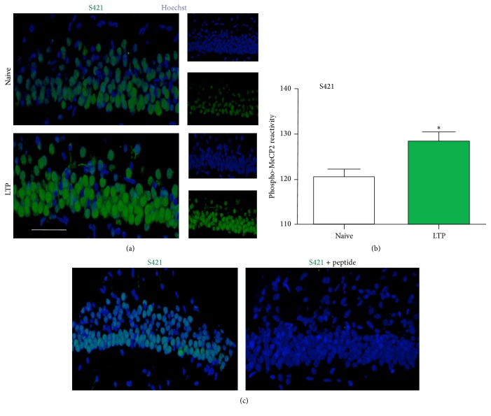
Figure 6.
The phosphorylation of MeCP2 at S421 at two hours after tetanic stimulation. (a) Representative immunostaining for phospho-S421 MeCP2 (green) and the nuclear marker Hoechst 33342 (blue) in the hippocampal CA1 region in naïve (number of nuclei = 136) and tetanized (number of nuclei = 164) slices at two hours after TBS. Calibration bar, 50 μm. (b) Quantification of phopho-MeCP2 reactivity for data such as that shown in (a). ∗ p < 0.05, using Student's t-test. (c) Immunostaining for phospho-S421 MeCP2 (green) and the neuronal marker Hoechst 33342 (blue) in the hippocampal CA1 region in tetanized slices at two hours after TBS in the absence (right panel) or presence of treatment with the immunogen peptide (left panel) to which the antibody was generated.

