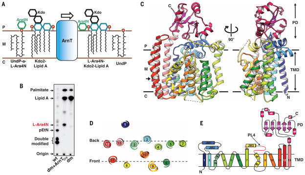
Fig. 1. Structure and function of ArnT from Cupriavidus metallidurans CH34.
(A) Schematic representation of the reaction catalyzed by ArnT. The sugar L-Ara4N is transferred from the carrier UndP to lipid A. 1 and 4′ phosphate positions on lipid A are marked. (B) Analysis of 32P-labeled lipid A species by TLC showing rescue of lipid A modification with L-Ara4N when ArnTCm is expressed in the ΔarnTΔeptA double-knockout E. coli strain (dm). The background E. coli strain (WD101; wt, wild type) has a pmrA constitutive phenotype (pmrAC), enabling it to synthesize L-Ara4N- and pEtN-modified lipid A and to exhibit resistance to polymyxins (8). (C) Crystal structure of ArnTCm. Two orthogonal views, perpendicular to the plane of the membrane, are presented in ribbon representation with rainbow coloring starting from the N terminus (blue) to the C terminus (purple). The approximate dimensions of the monomer are 55,79, and 42 Å (width, height, and depth). Membrane boundaries shown were calculated using the PPM server (21). Dashed lines represent missing segments in the structure. (D) Arrangement of TM helices in ArnTCm shown as a slice of the TM domain at the level indicated by the arrow in (C). (E) Schematic representation of the connectivity and structural elements of ArnTCm. P, periplasm; M, membrane (inner); C, cytoplasm; TMD:TM domain; PL4 (shown in red).

