
Figure 1. TLR11 −/− mice do not support S. Typhi replication.
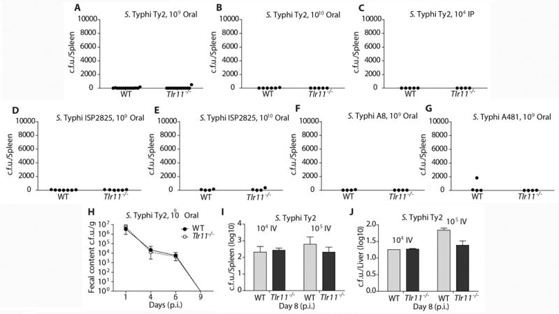
(A–G) Groups of sex- and age (6 to 10 weeks)-matched control and TLR11−/− mice were infected with the indicated S. Typhi strains. Infection routes and doses are indicated in the figure panels. Shown are c. f. u. 24 days after infection obtained from spleens of animals infected with S. Typhi Ty2 (A–C), ISP2825 (D and E), A8/14353363984-6 (F) and 3290A481 (G). (H) Groups (n = 5) of male and female mice aged 9 to 14 weeks were inoculated via oral gavage with approximately 1×10¹⁰ CFU of S. Typhi Ty2 in a volume of 0.2 ml. Fresh fecal pellets were collected at indicated time points. (I and J) Mice infected intravenously infectd with the indicated dozes of S. Typhi Ty2 and the c. f. u. in spleens (I) and livers (J) enumerated 8 days after infection. See supplemental experimental procedures for information on each laboratory’s experimental contributions.