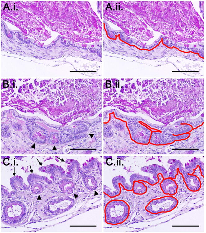
Figure 2. Architecture of TEI produced from enteroid seeding.

Shown are representative images demonstrating: A.i) simple columnar epithelium, B.i) flat simple columnar epithelium with underlying crypt domains (arrowheads), and C.i) crypt domains (arrowheads) with overlying blunted villi (arrows). A.ii-C.ii) Respective images of the varying types of neomucosa with the basement membrane traced in red to demonstrate how the neomucosa was quantified. All images are stained PAS. The bright pink staining in the lumen represents retained cellular debris and mucin. Scale bars = 100 μm.
