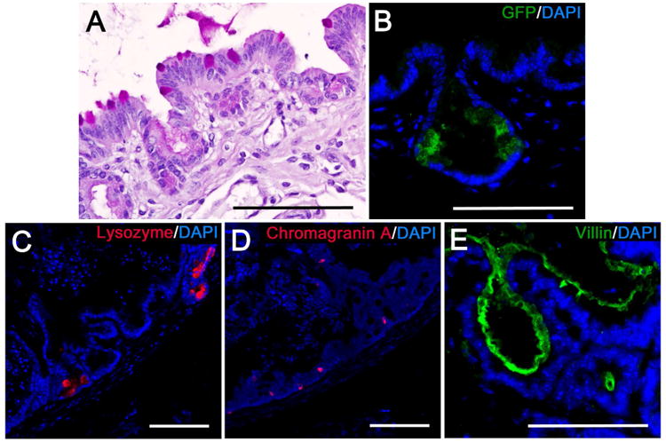
Figure 5. TEI contains ISCs and differentiated intestinal epithelial cells.

Shown are representative images demonstrating: A) PAS staining to identify goblet cells as noted by mucin filled vacuoles that stain bright pink, B) IF labeling for green fluorescent protein (GFP) (green) to identify LGR5+ ISC in crypt domains. C) IF labeling of lysozyme (red) to identify Paneth cells, D) IF labeling of chromagranin A to identify enterochromaffin cells (red), E) IF labeling of villin in the brush border to identify enterocytes (green). For all IF images, nuclei labeled with DAPI are represented in blue. Scale bars = 100 μm.
