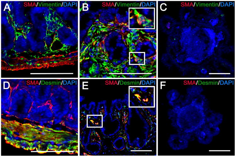
Figure 6. TEI contains ISEMFs and smooth muscle cells.

Shown are representative images from native intestine (A, D), TEI (B, E), and enteroids (C, F) that have been subjected to IF labeling for the identification of ISEMFs and smooth muscle cells. A-C) ISEMFs are identified by co-localization of labeling for smooth muscle alpha actin (SMA) (red) and vimentin (green). D-F) smooth muscle cells are identified by co-localization of labeling for SMA (red) and desmin (green). A) Native intestine shows strong SMA and vimentin co-localization in the submucosal layers only, indicative of ISEMFs. There is some vimentin staining in the muscularis that occurs in the mesenchymal stroma between the longitudinal and circular layers. B) TEI demonstrates the same SMA and vimentin co-localization surrounding the crypt domains. C) SMA and vimentin are not evident in enteroids. D) Native intestine shows strong SMA and desmin co-localization in the muscularis, indicative of smooth muscle cells. There is SMA labeling in the submucosa without desmin co-localization, consistent with ISEMFs. E) TEI demonstrates similar SMA and desmin co-localization surrounding all areas of the epithelium, however, it appears scattered and less organized than that observed in native intestine. F) SMA and desmin are not evident in enteroids. Scale bars = 100 μm.
