Fig. 2.

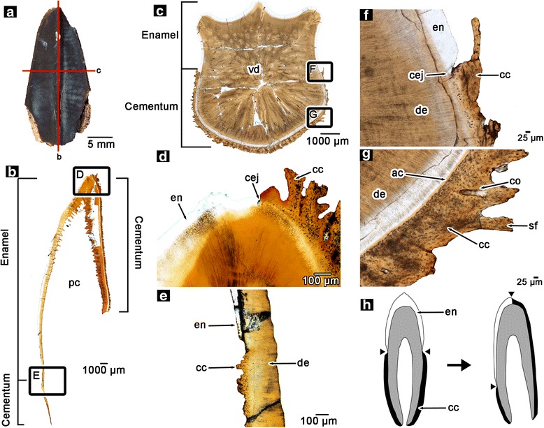
Histology of hadrosaurid teeth. a partial hadrosaurid tooth showing planes of section (ROM 58630). b isolated wholeview image of a coronal section through an in situ, unerupted maxillary tooth of a hadrosaurid (ROM 00696). c isolated wholeview image of a transverse section through an in situ, erupted maxillary tooth of a hadrosaurid (ROM 59042). d closup image of the apex of the tooth in (b). e closeup image of the base of the tooth in (b). f closeup image of the cemento-enamel junction of the tooth in (c). g closeup image of the cementum of the tooth in (c). h illustration of the anatomical differences between a generic amniote tooth (left) and a hadrosaurid tooth (right). Hadrosaurid teeth exhibit a displacement of the cementum and enamel relative to other amniotes. ac, acellular cementum; cc, cellular cementum; cej, cemento-enamel junction; co, cementeon; de, dentine; en, enamel; pc, pulp cavity; sf, Sharpey’s fibers; vd, vascular dentine
