Fig. 5.

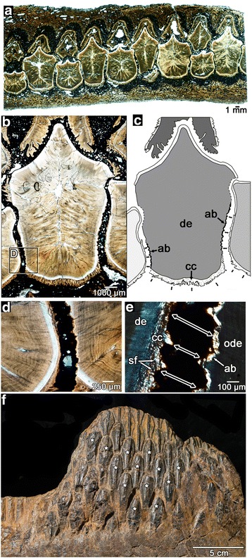
Tooth-to-tooth attachment within the hadrosaurid dental battery. a wholeview image of a transverse section through a maxillary battery, near the occlusal surface (labial is towards the top). Note that none of the teeth in the section make hard-tissue contacts to adjacent teeth. b closeup image of three neighbouring teeth in the same transverse section of a partial maxilla (ROM 59042). c illustration of (b) showing positions and orientations of periodontal ligament connections (black arrows) between adjacent teeth. Lighter shades of grey indicate younger relative ages for teeth, determined by partial resorption of older neighbouring teeth. Dashed lines indicate tooth resorption. d magnified image of (b) showing partial resorption of an older tooth (right) by a younger tooth (left). e magnified cross-polarized image of (d) showing the development of a thin layer of alveolar bone along the partially resorbed root of the older tooth. The younger tooth developed a periodontal ligament connection (black arrows) to the older tooth as indicated by the presence of Sharpey’s fibers. f a partial dentary battery of Edmontosaurus (ROM 00620) showing several teeth that have moved out of position post-mortem (asterisks), which supports the presence of a soft tissue connection between these teeth in life. ab, alveolar bone; ab/rc, alveolar bone (possible repair cementum); cc, cellular cementum; de, dentine; en, enamel; ode, dentine of older tooth; sf, Sharpey’s fibers
