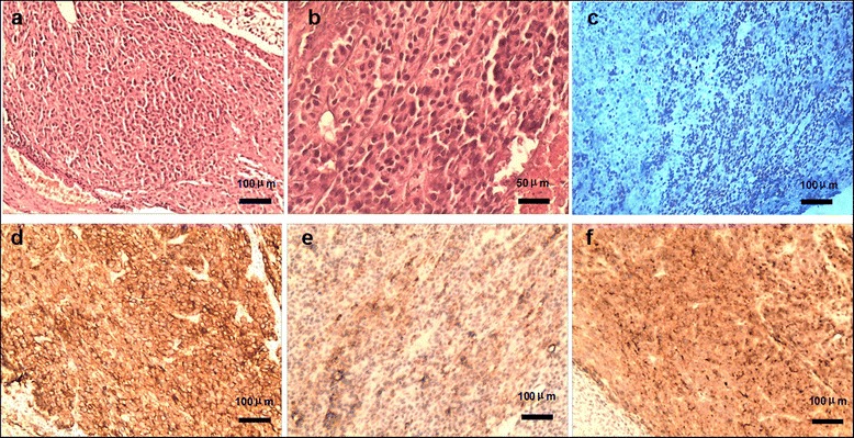
Fig. 3.

Pathological diagnosis of ectopic hepatocellular carcinoma. a HE staining, cancer cell nest with pseudoglandular structure and focal necrosis area (100×). b HE staining, morphologically typical hepatocellular carcinoma cells with polygonal shape, eosinophilic cytoplasm and big anachromasis nucleus. c Ki-67, positive in nucleus (60–70 %, 100×). d CK18, positive on membrane (++, 100×). e Hepatocyte, positive in cytoplasm (+, 100×). f Glypican-3, positive in cytoplasm (++, 100×)
