Fig. 3.

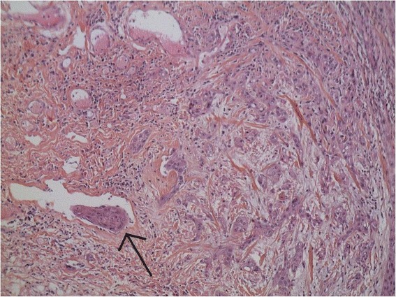
Fixed HE-stained pathology of excised tissue (original magnification 20×). The tissue corresponds to an infiltrative poorly-differentiated squamous cell carcinoma. Arrow indicates an embolus in the vessel

Fixed HE-stained pathology of excised tissue (original magnification 20×). The tissue corresponds to an infiltrative poorly-differentiated squamous cell carcinoma. Arrow indicates an embolus in the vessel