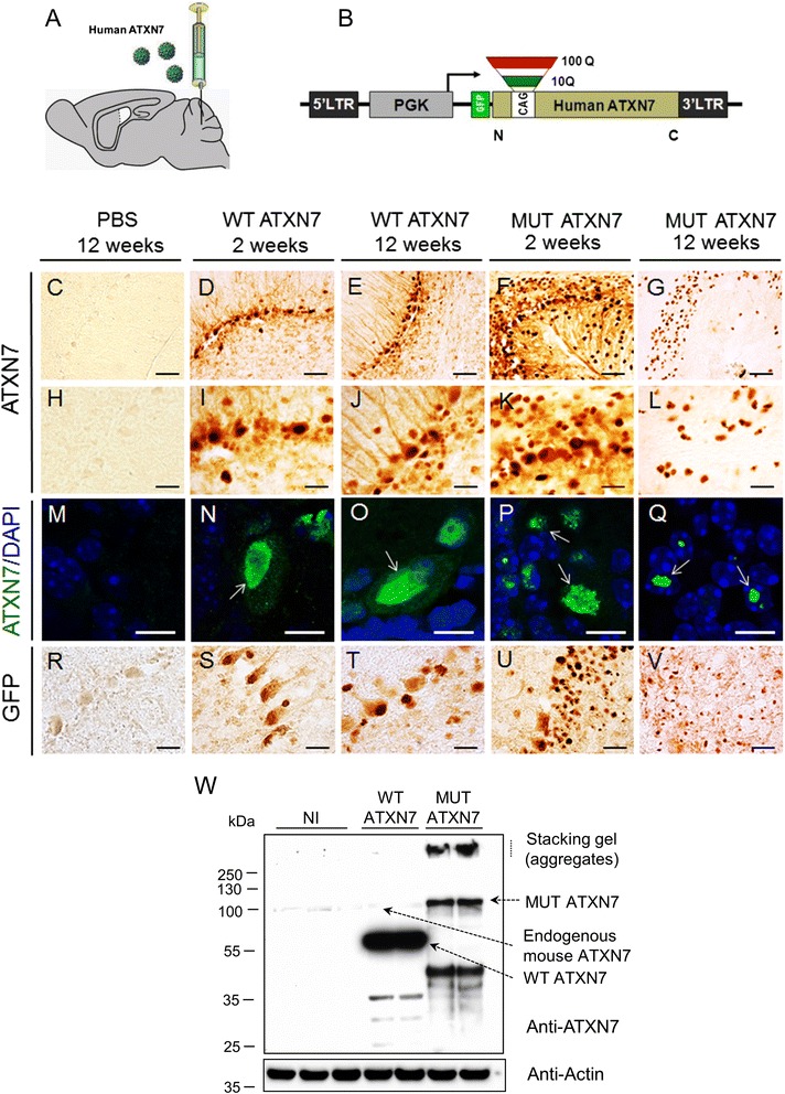
Fig. 1.

Lentiviral-mediated overexpression of truncated human WT ATXN7 and MUT ATXN7 in the mouse cerebellum. a Protocol: lentiviral vectors (LV) encoding truncated human WT ATXN7 or MUT ATXN7 were injected sterotactically into the mouse cerebellum. The animals were sacrificed at 2 and 12 weeks post-injection. b Schematic representation of the LV encoding truncated (amino acids 1–230) wild-type (WT ATXN7; 10 CAG repeats) or mutant (MUT ATXN7; 100 CAG repeats) human ATXN7 (ATXN7) and a GFP tag, under the control of the phosphoglycerate kinase-1 (PGK-1) promoter. c–l and (r–v) Immunohistochemistry and (m–q) immunohistofluorescence in cerebellum. Truncated WT ATXN7 was mainly nuclear and diffuse in PCs at 2 (D, I and N) and 12 weeks post-injection (E, J and O) (arrows), whereas MUT ATXN7 was nuclear and diffuse at 2 weeks (F and K), with small intranuclear aggregates in PCs (arrows in P) (laser confocal microscopy: ATXN7 in green; DAPI stain, blue). MUT ATXN7 forms robust intranuclear inclusions in the GCL at 12 weeks (G, L and arrows in Q). Immunohistochemical labeling with anti-GFP showed similar results (R-V). No transgenic ATXN7 immunoreactivity was observed in mice injected with PBS (C, H and M). w Representative western-blot of cerebellar lysates: overexpression of truncated WT ATXN7 and MUT ATXN7, 2 weeks post-injection, probed with an anti-ATXN7 antibody that recognizes both endogenous mouse and human ATXN7. Non-injected cerebella (NI) were used as control: human WT ATXN7 expression was ~30-fold higher than endogenous mouse ATXN7 (analysis by optical densitometry). ATXN7-positive aggregates were retained in the stacking gel (SDS-insoluble fraction) and a robust smear of putative cleavage fragments was observed in MUT ATXN7 but not in WT ATXN7-injected animals. Bars: C–G: 50 μm; H–L: 20 μm; M–Q: 10 μm; R–V: 20 μm
