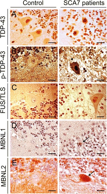
Fig. 8.

Immunoreactivity of TDP-43, FUS/TLS, MBNL1 and MBNL2 in the cerebellum of control and SCA7 patients. Representative immunohistochemically labelled cerebellar sections (counterstained with hematoxylin) from a SCA7 patient and a control with no neurological disease. a TDP-43 immunoreactivity was increased in the nucleus of PCs in the cerebellum of a SCA7 patient compared to control PCs where it remained diffuse with few TDP-43 dots. b Phosphorylated TDP-43 was strongly labeled in the nucleus of a PC of a SCA7 patient; only rare nuclear granules were observed in the nucleus of control PCs. c FUS/TLS shows increased immunoreactivity with FUS/TLS-positive small dots in the nucleus of a PC in a SCA7 patient compared to a control PC where FUS/TLS was almost not detectable in the nucleus. d Nuclear accumulation of the MBNL1 protein was higher in an atrophic PC in a SCA7 patient compared to PCs from a control. e MBNL2 diffuse immunoreactivity is increased in the nucleus and cytosol of a SCA7 PC, compared to control PCs where MBNL2 immunoreactivity is low in the cytoplasm and almost undetectable in the nucleus. Bars: 20 μm
