Figure 5.
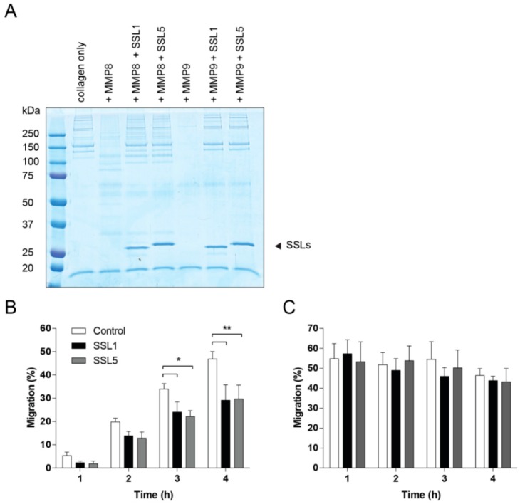
SSL1 and SSL5 limit neutrophil migration through collagen. (A) MMP-mediated collagen degradation was visualized using SDS-PAGE. Collagen (0.5 mg/mL) was incubated overnight with MMP8 and MMP9 (10 µg/mL) with and without the SSLs (10 µg/mL). Samples were loaded on gel and visualized using Instant Blue; (B,C) Migration of neutrophils was assessed in presence (B) or absence (C) of a collagen gel layer in the upper compartment of a Transwell system. Fluorescently labeled neutrophils, untreated (white bars) or treated with 10 µg/mL SSL1 (black bars) or SSL5 (gray bars), and placed on top of the collagen layer, were allowed to migrate for 1, 2, 3 and 4 h through the gel towards the chemo-attractants fMLP and LTB4 present in the lower compartment. Migration was monitored every hour by measuring the amount of fluorescence present in the lower compartment. Data points represent mean plus SE for at least three independent experiments. * p ≤ 0.05, ** p ≤ 0.01.

