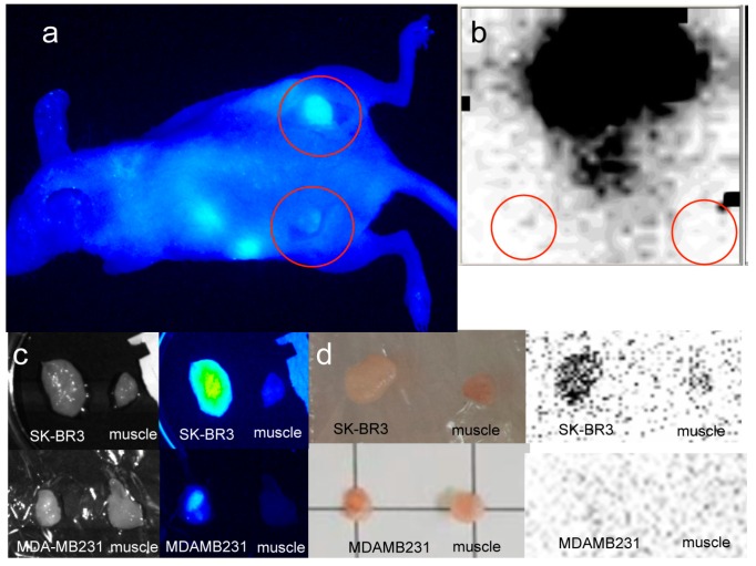
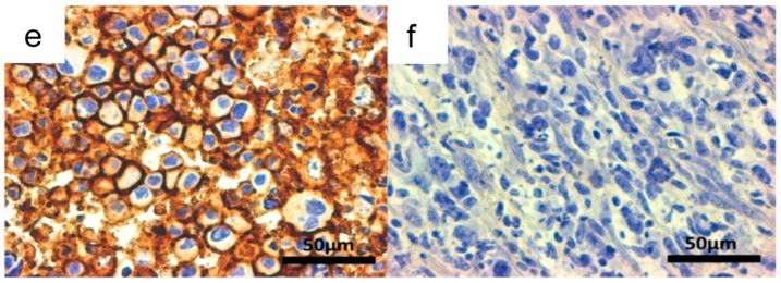
Figure 7.
Xenografted tumors image with direct-99mTc-chelated dual-imaging probe (red circles). A NIR imager reveals high fluorescence intensity in the SK-BR3 tumor (right dorsum, a) in contrast to the MDA-MB231 tumor (left dorsum, a) in a single mouse bearing both types of tumors in the direct-99mTc-chelated dual-imaging probe studies. The NIR image was taken after skin incision over the xenografted tumors. The tumor-to-muscle ratios of fluorescence intensity and radioactivity were higher in the SK-BR3 tumor than in the MDA-MB231 tumor. Specific tumor uptake is not confirmed by a small γ camera with acquisition time 30 s in the lower-body image (b). Excised SK-BR3 tumors exhibit a higher fluorescence intensity in a NIR imager image (c, upper right) and a higher radioactivity in an FLA-2000 image (d, upper right) than do muscle tissue and MDA-MB231 tumors (c, bottom right, d, bottom right). The size of the SK-BR3 tumor is 13 mm × 8 mm × 2 mm and the size of the MDA-MB231 tumor is 6 mm × 8 mm × 5 mm (c,d). HER2 immunohistochemistry reveals stronger positive stains in the SK-BR3 tumor cells (e) than in the MDA-MB231 tumor cells (f).


