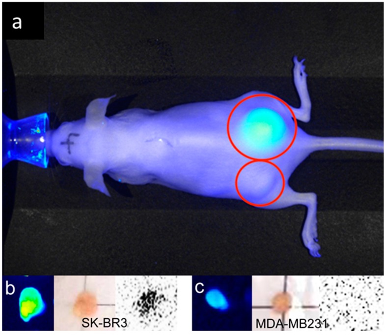
Figure 9.
Xenografted tumors image with 99mTc-diethylenetriamine pentaacetic acid (DTPA) dual-imaging probe (red circles). In vivo imaging of tumor xenograft mice with use of DTPA used dual-imaging probe. The NIR image showed a high-intensity fluorescence in the SK-BR3 tumor (right dorsum, a) in contrast to the MDA-MB231 tumor (left dorsum, a). Excised SK-BR3 tumors (b) exhibit a higher fluorescence intensity and radioactivity in contrast to the MDA-MB231 tumor (c). The size of the SK-BR3 tumor is 11 mm × 8 mm × 2 mm and the size of the MDA-MB231 tumor is 6 mm × 5 mm × 5 mm (b,c).

